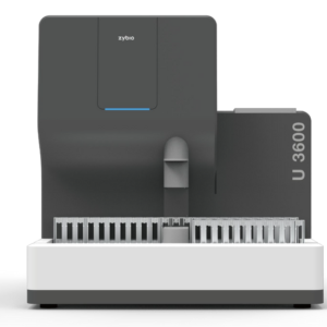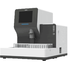


Automatic urine sediment analyzer is a device with the function of analyzing urine sediment, indicators such as: Red blood cells (RBC), white blood cells (WBC), white blood cell mass (WBCC), phagocytosis (PHCY ), squamous epithelial cells (SQEP), non-squamous epithelial cells (NSE), budding yeast (BYST), yeast with pseudohyphae (HYST), bacilli (BACI), suspected coccidiosis (SUCO), spermine (SPRM), mucoid (MUCS), clear cast (HYAL), Unclassified cast (UNCC), fatty, amorphous (AMOR), crystalline magnesium ammonium phosphate (MAPH), calcium oxalate monohydrate (COM), calcium oxalate dihydrate crystals (COD), uric acid crystals (URIC), ammonium urate crystals (AUCR), tyrosine crystals (TYRO), calcium phosphate crystals (CAPH), undissolved crystals type (UNCX) and unclassified (UNCL) and derivative and osmotic parameters, the machine has high capacity, convenient operation, accurate identification results.
Product features
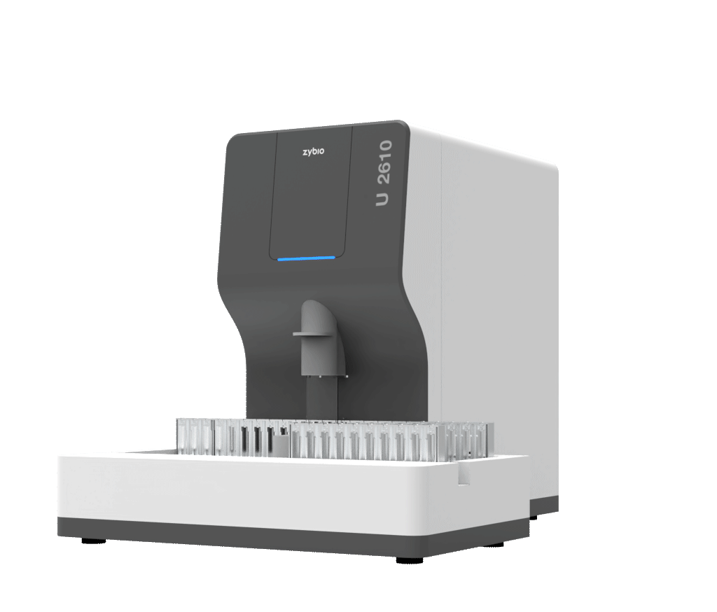
- Slim design: easy-to-use touch screen, intuitive navigation,
- Convenient operation: Samples are mixed automatically, no need for centrifugation, emergency samples are prioritized
- Accurate results: High definition images increase detection rate, no overlap, no agglomeration, no change in deposit shape, large, reliable database
- Capacity: 150 tests/hour
Operating principle:
The automatic urine sediment analyzer applies the principle of flow microscopic imaging. The device’s hydrodynamic system consists of a specially designed flow cell with a thin layer structure. After aspirating the sample, the sample enters the flow cell and injects the outer liquid solution (sheath fluid) into the flow cell, surrounded by the outer liquid solution (sheath fluid), the sample enters the thin layer structure . The cells in the sample flow sequentially through the thin layer structure. The deposit area is exposed to a high-frequency light source shaped by the illumination component, while the camera captures images of the deposit at the same frequency and transmits the deposit image to computer software. The computer software analyzes the target sediment images, analyzing the morphology, statistics, frequency domain, and texture of the target image, normalizing these features as input to the classifier. After classifying and calculating using the computer software’s identification algorithm, the urine sample will be discharged into the liquid waste container.